What is Twin-to-twin Transfusion Syndrome?
Twin-to-twin transfusion syndrome is a specific disease unique to multiple pregnancies in which two or more foetuses share the same placenta. In this condition, the problem derives from an abnormally unbalanced flow of fluids between the foetuses. While one of the foetuses receives less blood and subsequently grows and urinates less, the other foetus receives more blood, urinates more and is usually bigger. The problem can be resolved with minimally invasive surgery that separates the foetuses into two individual placentae. The syndrome usually appears between weeks 16 and 25 of pregnancy.
Twin-To-Twin Transfusion Syndrome explained in first person
Fetoscopy is a minimally invasive technique that, definitely, separates two foetuses in two independent placentas.
It seems worse than it is, because if you follow your doctor’s recommendations, everything will be fine. Don’t be scared. It seems worse than it really is. It’s hard, but it’s worth it.
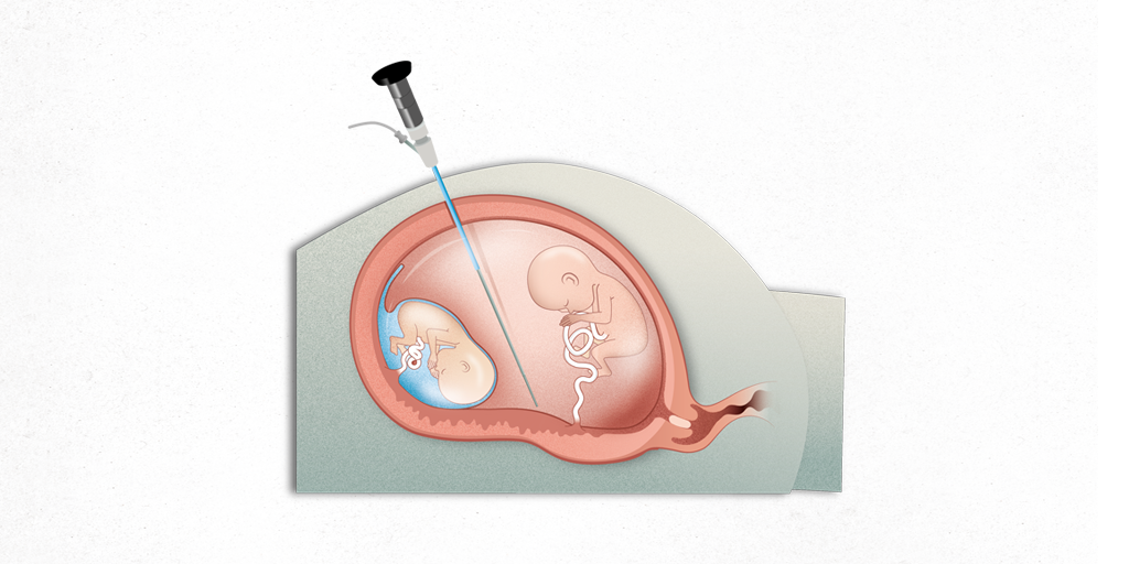
Twin-to-twin transfusion syndrome (TTTS) is a specific complication unique to multiple pregnancies that have just a single placenta (monochorionic twins). It develops when there is an unbalanced transfer of blood from one foetus to the other by means of abnormal, interconnecting blood vessels (anastomoses) running through the shared placenta. The foetus transferring the blood is known as the “donor”, while the foetus accepting it is called the “recipient”.

What is a monochorionic twin pregnancy?
One of the most important aspects in the management of multiple pregnancies is the diagnosis of chorionicity, in other words, identifying how many placentae are present.
Twin pregnancies can be classified as depending on the number of placentae:
- one shared placenta (monochorionic)
- two independent placentae (dichorionic)
Chorionicity must be diagnosed before week 15 in order to consider the result reliable. Monochorionic pregnancy is diagnosed by identifying the T sign in ultrasound images (direct connection between the two amniotic membranes without any interposing placenta); this is possible in almost 100% of cases at this gestational age. In a dichorionic pregnancy the chorion (placenta) extends between the two amniotic sacs, observed as a λ sign (lambda) in ultrasounds.
Monochorionic pregnancies are further categorised as:
- two amniotic sacs (diamniotic)
- both foetuses share a single amniotic sac (monoamniotic)
Is twin-to-twin transfusion syndrome very common?
There is one monochorionic (MC) twin pregnancy in every 250 pregnancies, they correspond to around 20% of all twin pregnancies and 70% of monozygotic pregnancies (those derived from a single ovum).
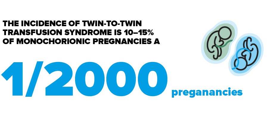
The incidence of twin-to-twin transfusion syndrome is 10–15% of monochorionic pregnancies and so it affects approximately 1 in every 2,000 pregnancies.
Types of twin-to-twin transfusion syndrome
The classification is based on ultrasound data and categorises the severity of the syndrome. It is divided into 5 stages (I–V). In stages I and II the foetuses do not present any severe cardiovascular alterations. More severe alterations start to develop in stage III and by stage IV one of the foetuses will present advanced heart failure.
- Stage I. There is amniotic fluid imbalance between the donor and recipient (polyhydramnios/oligohydramnios). The bladders also present different sizes; the recipient has a full and swollen bladder throughout the ultrasound examination, whereas the donor has a smaller bladder.
- Stage II. The donor foetus’ bladder is no longer visible.
- Stage III. This is a more advanced stage of the disease in which alterations appear in the recipient and/or donor foetus’ circulation; these are the result of haemodynamic changes secondary to an increase (hypervolaemia) or decrease (hypovolaemia) in the total volume of blood circulating in each foetus. The most common alterations are:
- Absent or reversed end-diastolic flow in the donor and/or recipient’s umbilical artery.
- Absent or negative A wave in the ductus venosus (usually in the recipient).
- Umbilical vein with pulsatile flow (usually in the recipient).
- Stage IV. Hydrops in either of the foetuses (accumulations of fluid in two or more foetal compartments, e.g., subcutaneous, pleural effusion, ascites, pericardial effusion).
- Stage IV. Intrauterine death of one or both twins.
Substantiated information by:


Published: 20 February 2018
Updated: 8 July 2025
Subscribe
Receive the latest updates related to this content.
Thank you for subscribing!
If this is the first time you subscribe you will receive a confirmation email, check your inbox
