Diagnosis of Lymphoma
It is essential to carry out a biopsy on one of the enlarged lymph nodes whenever lymphoma is suspected. Considering each node gives the same result, doctors tend to select whichever is most accessible. Hence, if a patient’s neck and abdominal lymph nodes are inflamed, then the neck will be selected because the procedure is more straightforward and recovery is quicker.
However, if only abdominal nodes, or those in the chest, are effected, then there is no choice but to employ more complicated surgical techniques, such as mediastinoscopy (a surgical procedure used to examine the mediastinum, the space which separates the lungs located behind the sternum and in the centre of the thorax) or laparoscopy (exploration or examination of the abdominal cavity by introducing a laparoscope through a small incision).
Surgery used for lymphomas
Surgery only serves to recover a sample and confirm the diagnosis under a microscope. Hence the surgeon will not try to remove “all of the lymph nodes possible”, as they do with other cancers, such as breast cancer, cancer of the larynx, etc.; extracting just one node will be enough.
In some cases, the disease does not appear in a lymph node and so this may complicate the diagnosis. It sometimes originates in the spleen (which is also a lymphatic organ), in the digestive system (stomach, intestine, which also feature lymphatic tissues), the tonsils, or even inside bones (bone marrow).
Study to determine the degree of spread
Once the diagnosis of lymphoma has been confirmed, doctors then need to know how much it has spread. This mainly involves two tests:

Positron Emission Tomography (PET-CT) or Computed Tomography (CT). These are radiological tests used to determine the size and location of all the body’s lymph nodes, as well as the rest of the organs (heart, lungs, liver, kidneys, etc.).

Bone marrow biopsy. This test is conducted because lymphomas often spread to the inside of bones.
Bone marrow biopsy
A bone marrow biopsy is often carried out in patients diagnosed with lymphoma to discover whether the disease has spread to the bones as this is important when selecting the treatment.
The test involves the extraction of a small core of pelvis bone (the hip bone). The core can be extracted from the rear of the bone but it is also often collected from the front.
Patients must inform the medical team if they are taking any medications, such as aspirin, antiplatelet agents (clopidogrel) or anticoagulants (Sintrom®). They must also mention any history of allergies to anaesthetics.
Degree of spread
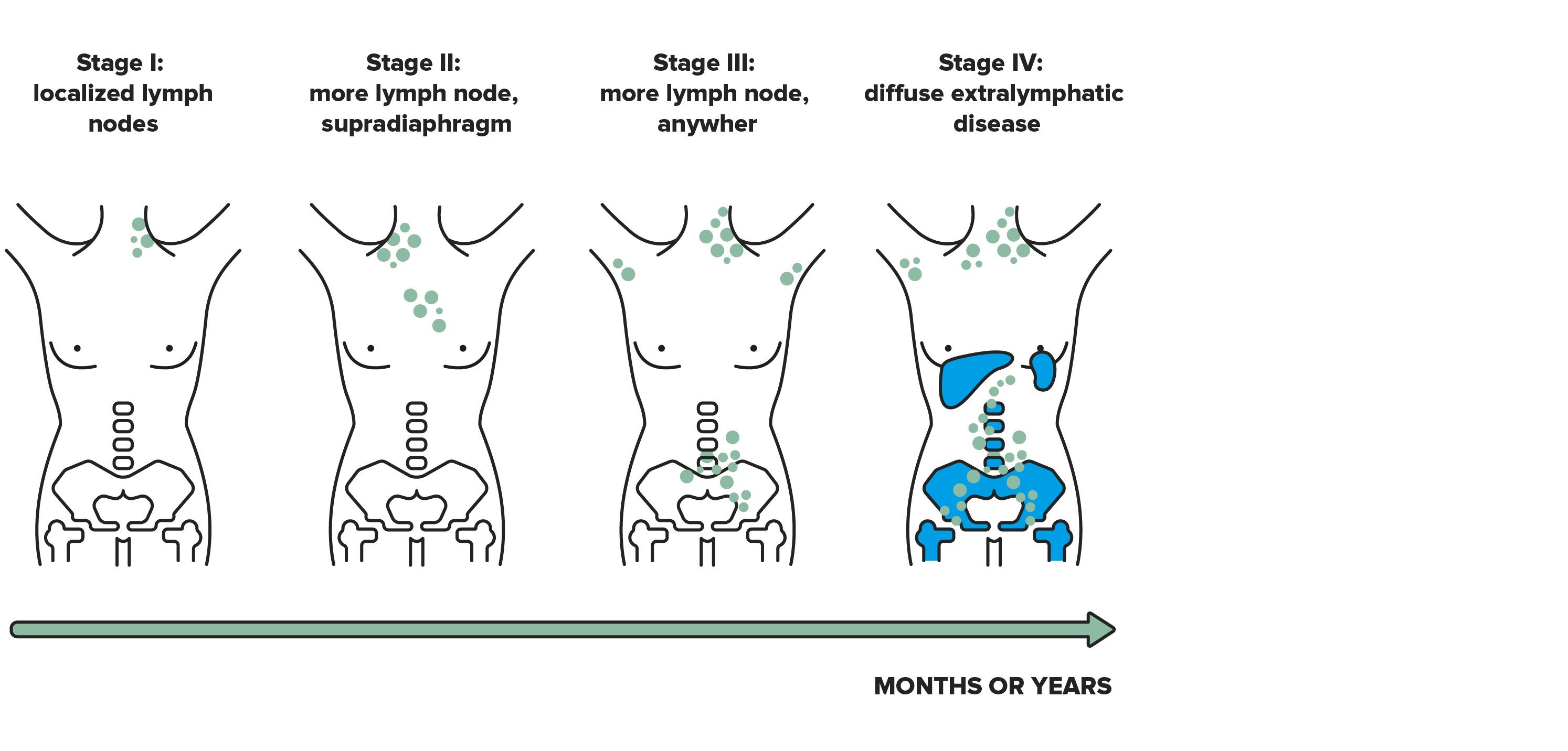
The results of these tests (PET-CT or CT, and bone marrow biopsy) can be used to determine the cancer’s degree of spread. This is classified according to the Ann Arbor staging system.
Stage I. Only one lymph node is affected, the cancer is confined to a single region.
Stage II. Two lymph nodes located close to each other are affected (they are either all above or all below the diaphragm, the muscle separating the thorax from the abdomen).
Stage III. More than one lymph node is affected in different areas of the body (on both sides of the diaphragm).
Stage IV. There is diffuse involvement in areas other than the lymph nodes or spleen (bone marrow, liver, lungs, pleurae, kidneys).
These stages are further subdivided into A or B in function of whether or not the patient presents symptoms associated with lymphoma (fever, sweats, weight loss, etc.).
Substantiated information by:


Published: 20 February 2018
Updated: 20 February 2018
Subscribe
Receive the latest updates related to this content.
Thank you for subscribing!
If this is the first time you subscribe you will receive a confirmation email, check your inbox


