What is Evoked Potentials test?
Evoked potentials are diagnostic tests that evaluate how the nervous system responds electrically when stimulated by a light source, sound or electrical current.
The brain’s response to these stimuli is recorded by electrodes, which measure both the response speed (how long it takes for the signal to reach the brain) and its magnitude (how much electrical activity is generated).
What types of Evoked potentials exist?
There are four types of evoked potentials:

Visual evoked potentials (VEPs). These assess the functioning of the pathway that transmits visual information to the brain; the pathway runs from the retina to the rear part of the brain (the occipital lobe).
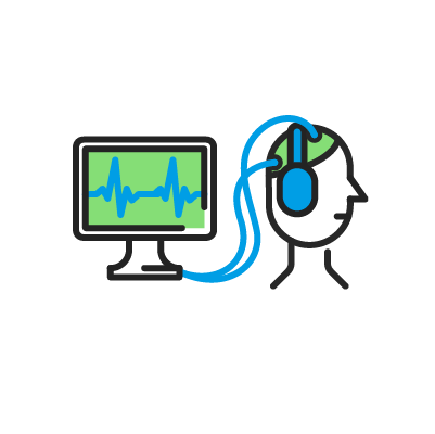
Brainstem auditory evoked potentials (BAEPs). These assess the functioning of the pathway that carries auditory information to the brain; this runs from the ear to the lateral part of the brain (the temporal lobe).
Somatosensory evoked potentials (SSEPs). These evaluate the functioning of the pathway that transmits sensory information—such as touch, vibration or joint movement—to the brain; this pathway runs from the nerves in the arms and legs to the brain’s parietal lobe.
Motor evoked potentials (MEPs). These evaluate the functioning of the motor pathway, which transmits signals from the brain to the muscles to enable movement; it runs from the frontal lobe to all the body’s muscles.
When is this test necessary and what diseases can it diagnose?
This test determines whether a neural information pathway is functioning properly or not.
Impaired functioning of one of these pathways may result in vision or hearing loss, numbness, difficulty moving the arms or legs or problems with walking.
Each type of evoked potential assesses a different neural pathway, so it is possible to record the activity of:
-
The optic nerve in demyelinating diseases, such as multiple sclerosis.
-
The auditory nerve, which may be affected by intracranial lesions or those within the auditory canal, such as meningiomas or schwannomas.
-
Sensory and motor pathways in brain or spinal cord injuries that are close to these pathways.
-
All pathways (visual, auditory and sensory) if there is a discrepancy between clinical symptoms and findings from other tests (e.g., MRI, audiometry or optical coherence tomography) or for individuals who are unable to cooperate (e.g., young children or patients in a coma).
-
All pathways during surgery on brain and spinal cord lesions located near these pathways, with the aim of preventing severe, irreversible injury during the procedure.
It can also help determine neurological prognosis in individuals who have experienced cerebral hypoxia after a cardiorespiratory arrest.
How should I prepare for the procedure?
-
There is no need to fast.
-
Continue taking your usual medication.
-
Do not use any creams or lotions on the body.
-
Do not use hair sprays or styling products, like gel or cream, beforehand, as they can make it difficult for the electrodes to adhere to the scalp properly, causing them to fall off during the test.
-
Avoid consuming caffeine or other stimulants, so you are relaxed during the test.
-
Inform healthcare professionals if you have a pacemaker or other electronic device or neurostimulator fitted for pain, epilepsy or Parkinson’s disease (e.g., spinal cord or deep brain stimulation).
How is the test performed?
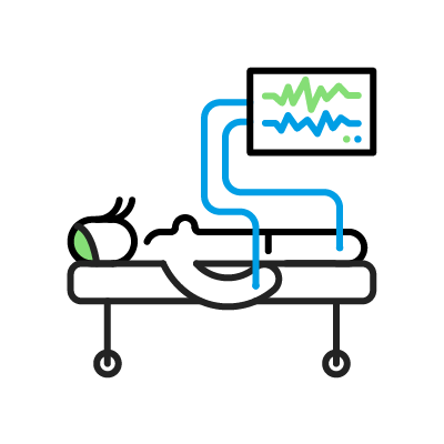
Electrodes or small sensors are placed on the head (scalp) and, in some cases, also on the arms or legs, after the skin has been gently exfoliated with a gel. The electrodes are attached using a creamy paste and do not pierce the skin. These sensors record the electrical activity produced by the brain or its nerves.
Then, visual, auditory or electrical stimuli are applied, depending on the type of test required.
For visual evoked potentials, patients sits in a chair and look at the centre of a screen positioned in front of them. The screen displays black-and-white squares that change intermittently. If patients are unable to fix their gaze on one point, they lie on an examination table and a flashing white light is applied using a lamp. Each eye is tested separately by covering the other eye with a patch.
For auditory evoked potentials, the patient lies on an examination table with a pair of headphones. A repetitive sound is delivered in one ear and background noise is emitted in the other.
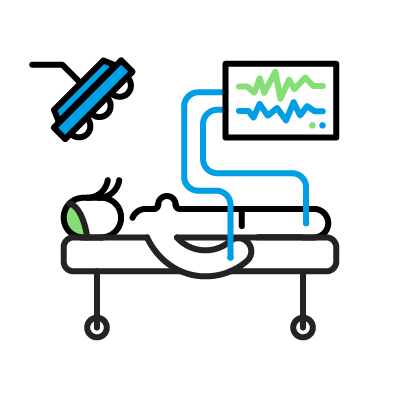
For somatosensory evoked potentials, the patient lies on an examination table and a small electrical current is used to stimulate the median nerve at the wrist and the posterior tibial nerve near the ankle.
The patient will feel a tingling or pulsating sensation, like a slight pinprick, which may cause the muscle of the thumb or big toe to move repeatedly.
Motor evoked potentials are performed exclusively in the operating room or Critical Care Unit. This is because a higher-intensity electrical stimulus is applied, and the test must be carried out under anaesthesia.
The recordings are stored digitally for later review by a healthcare professional, who prepares a report with the results.
An evoked potential test is painless, safe and non-invasive. The most important thing is to remain relaxed and not move during the procedure.
The average duration of the test is about one and a half hours, although this may vary depending on whether one or more types of evoked potentials are requested and on the patient’s level of relaxation.
Who performs the test?
The test is performed by a technician with specific training in neurological diagnosis. The results are reviewed by a specialist healthcare professional.
What are possible complications?
In general, this test causes no problems, with any possible effects being minimal and temporary.
-
Mild discomfort (similar to a pinprick or brief muscle spasm) may be felt during electrical stimulation in the somatosensory evoked potential test; but this stops when the stimulation ends. The stimulation is delivered at the lowest intensity necessary for accurate testing, and the technical staff will inform you where and when the electrical stimulation will be felt.
-
Minor involuntary movement may be experienced when electrical stimuli are applied during a somatosensory evoked potential test as the muscle moves slightly. However, this is usually not bothersome and is not dangerous.
-
Visual fatigue or eye strain may be experienced for a few minutes after repeated light stimulation.
-
Although very rare, some people with sensitive skin may suffer an allergic reaction or mild skin irritation to the conductive paste or exfoliating gel used with the electrodes.
There is no risk of injury to the brain or to the optic, auditory or peripheral nerves of the arms or legs. If any discomfort occurs, the test is stopped immediately.
Related contents
Substantiated information by:
Published: 10 February 2026
Updated: 10 February 2026
Subscribe
Receive the latest updates related to this content.
Thank you for subscribing!
If this is the first time you subscribe you will receive a confirmation email, check your inbox