Diagnosis of Age-related Macular Degeneration
The eye specialist may request a series of painless useful tests for the diagnosis of AMD. These tests will reveal if there is any damage in your eyes and/or impaired vision.

Visual acuity test. Measures your ability to see objects at different distances.

Amsler grid. A simple test you can use to check yourself is by using a grid with a dot in the centre. If the image is distorted (wavy lines, vertically or horizontally), irregular or you see any blind spots (scotomas) you should seek professional advice.

Fundus examination with pupil dilation. To examine the fundus (macula and optic nerve), the doctor applies drops to dilate the pupil. The effect of the drops can last from 4 to 12 hours, depending on the patient’s susceptibility.

Fundoscopy or ophthalmoscopy. This is performed with an optical instrument that emits a beam of light, so the examiner can visualise the macula, retina, blood vessels and optic nerve.

Optical coherence tomography (OCT). Imaging technique that uses light waves to take photographs of your retina and therefore measure and map theis thickness and features (shape and size).

Autofluorescence. Autofluorescence is a technique used to determine the area affected by retinal pigment epithelium (RPE) atrophy in patients with advanced dry AMD.

Retinography. This is a colour photograph of the fundus. A special camera is used to view part or all of the fundus.

Fluorescein angiography and indocyanine green angiography. Tests that examine blood flow in the capillaries of the retina (fluorescein) or choroids (indocyanine green).
Why do they have to dilate my pupils?
Dilating the pupils is a very common procedure during eye examinations. It is a totally painless process that allows the ophthalmologist to examine the fundus of your eye and determine the condition of your retina and optic nerve. It helps in the diagnosis of various eye diseases that may otherwise go unnoticed during early stages: macular degeneration, retinal detachment, glaucoma, diabetic retinopathy, etc.
The pupils are dilated by placing a few drops in the eye which act on muscles around the pupil, thus producing the dilation.
The drops may cause a slight itchiness just after they have been applied, but it is only temporary and will disappear shortly. You have to wait for 20 to 45 minutes after the drops have been instiled before your eyes dilate fully and the ophthalmologist can start the eye exam.
It is important for patients to take into account that the effects of dilation eye drops can last for several hours after the eye exam. Specifically, between 4 and 12 hours, or sometimes even more. This will depend on the type of drops used and each patient’s sensitivity. People are normally more sensitive to light and have blurred vision during this period in which the eye drops continue to have an effect. Although the effects will gradually disappear as the hours pass, patients are advised to wear sunglasses after receiving dilation eye drops and should be accompanied when they go to the eye exam. You must never drive after having your pupils dilated.
The effects of dilation eye drops last for longer in children than they do in adults. Similarly, blurred vision and sensitivity to light typically last longer in children than in adults.
How to use an AMSLER grid

The Amsler grid test is a very straightforward and useful tool for assessing your central vision. This is because it can detect changes in the macula very early on that would otherwise go unnoticed.

Position your face approximately 30 cm from the paper or the screen

If you normally wear glasses or contact lens, put them on

Cover one eye
Stare at the point in the centre of the grid
Repeat the process for the other eye
After completing the test, ask yourself:
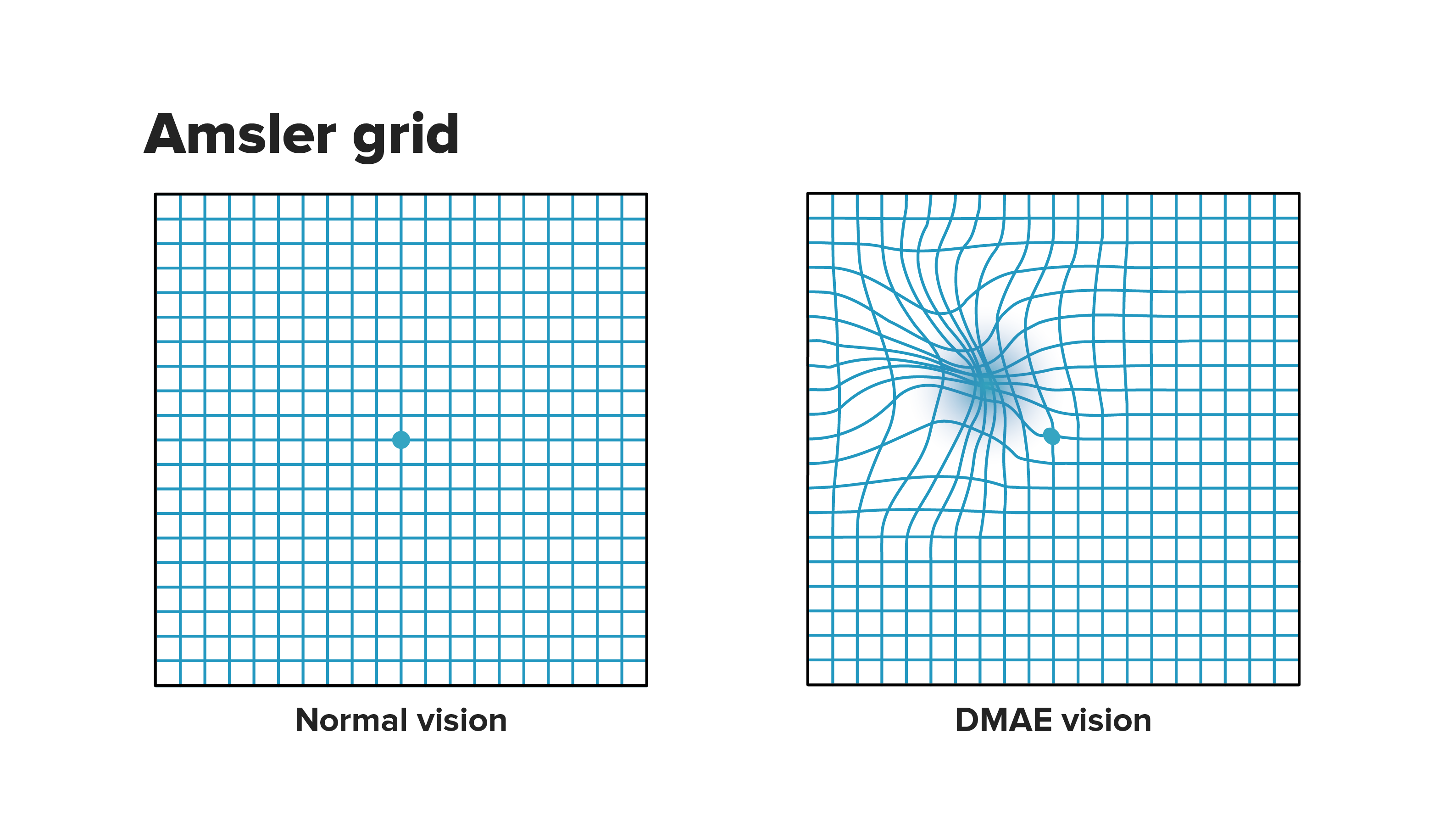
- Did all of the squares appear symmetrical?
- Did any lines appear to be twisted or bent?
- Did any of the lines appear wavy, blurred or disappear at any point?
If you answer “Yes” to any of these questions, then you should visit your ophthalmologist as soon as possible. Remember that this test only provides an indication and does not imply a diagnosis.
Do not leave it too late! It is important that you get your vision checked. Visit your ophthalmologist once a year and if you notice any changes in your vision visit your doctor.
Substantiated information by:


Published: 20 February 2018
Updated: 11 July 2025
Subscribe
Receive the latest updates related to this content.
Thank you for subscribing!
If this is the first time you subscribe you will receive a confirmation email, check your inbox


