

20 January 2026
PortalClinic home
Joint project with
BBVA Foundation website.This link opens in a new tab.
What types of Alopecia are there?
Reading time: 9 min
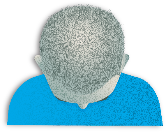
Androgenetic alopecia (male pattern)
What is male androgenetic alopecia?
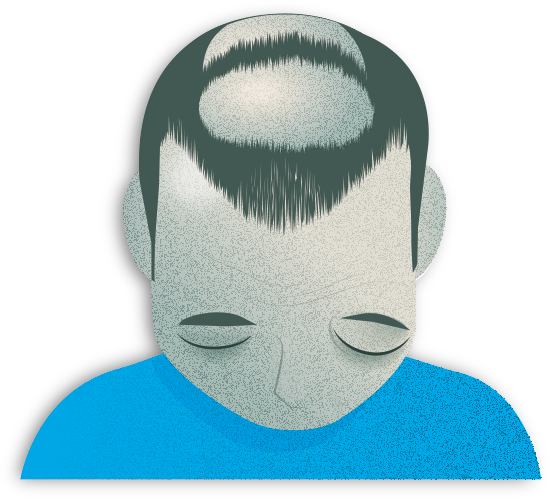
Male androgenetic alopecia is a type of non-scarring alopecia characterised by progressive hair thinning, particularly at the temples (frontotemporal regions) and the crown (vertex), as described in the Hamilton–Norwood classification. It is the most common form of alopecia in men, and it is estimated that more than 50% will develop this type of alopecia from around age 30, with its frequency increasing with age.
Signs and symptoms of male androgenetic alopecia
The signs of male androgenetic alopecia include progressive recession of the frontal hairline, hair thinning and reduced density in the upper scalp and crown. In more advanced stages, areas of marked hair loss appear in the frontal and superior regions, while hair is generally preserved on the sides and at the nape of the neck.
Causes of male androgenetic alopecia
Male androgenetic alopecia is caused by the action of male hormones (androgens—particularly dihydrotestosterone) on hair follicles in individuals with a genetic predisposition.
A family history is one of the main risk factors.
Diagnosis of male androgenetic alopecia
Male androgenetic alopecia is diagnosed through clinical examination by a specialist, based on observation of the scalp. Trichoscopy is used to confirm the diagnosis. This test allows assessment of variability in hair shaft thickness, an increased number of miniaturised follicles, reduced hair density and the presence of follicular units containing fewer hairs.
Although this type of alopecia is associated with hair ageing, its accelerated progression and its functional and aesthetic impact on the patient justify treatment.
Treatment of male androgenetic alopecia
Treatment includes topical or oral therapies that help slow hair loss and strengthen the hair (e.g. topical or oral minoxidil); medications that reduce the action of certain hormones (topical and systemic antiandrogens such as finasteride and dutasteride); as well as complementary therapies aimed at improving follicular quality (e.g., mesotherapy and platelet-rich plasma). In selected cases, hair transplantation may also be considered as a reconstructive option.
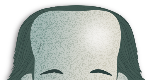
Androgenetic alopecia (female pattern)
What is female androgenetic alopecia?
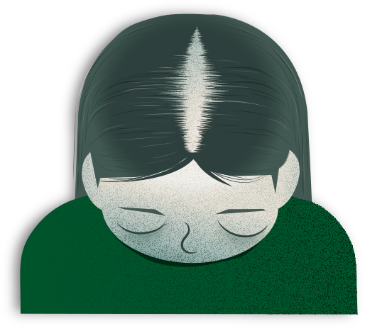
Female androgenetic alopecia is a type of non-scarring alopecia with typical symptoms of diffuse, progressive hair loss in the central region of the scalp. It generally preserves the frontal hairline and leads to the characteristic widening of the central parting line, as described in the Ludwig classification or the Sinclair scale.
The prevalence of female androgenetic alopecia increases with age, particularly after the age of 40 and following menopause. However, it may also occur in younger women, even from puberty.
Signs and symptoms of female androgenetic alopecia
Characteristic signs of female androgenetic alopecia are progressive hair thinning and reduced density in the upper scalp. Widening of the central parting line and increased visibility of the scalp are common, while the frontal hairline is usually preserved. Unlike male androgenetic alopecia, hair loss is more diffuse and rarely results in completely bald areas.
Causes of female androgenetic alopecia
Female androgenetic alopecia is influenced by genetic predisposition and hormonal factors. It is also associated with conditions such as polycystic ovary syndrome, thyroid disorders, iron deficiency and other nutritional deficiencies.
Diagnosis of female androgenetic alopecia
Female androgenetic alopecia is diagnosed through clinical examination by a specialist, based on observation of the scalp. Trichoscopy is used to confirm the diagnosis. This test allows assessment of variability in hair shaft thickness, an increased number of miniaturised follicles, reduced hair density and the presence of follicular units containing fewer hairs.
Treatment of female androgenetic alopecia
Treatment combines topical or oral therapies that help slow hair loss and strengthen the hair (e.g. topical or oral minoxidil) with antiandrogen therapy in women without contraindications and under medical supervision; correction of any existing deficiencies; as well as complementary therapies aimed at improving follicular quality (e.g., mesotherapy and platelet-rich plasma). In selected cases, hair transplantation may also be considered as a reconstructive option.
Alopecia areata
What is alopecia areata?
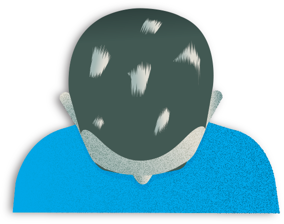
Alopecia areata is a type of non-scarring alopecia that presents as sudden, well-defined hair loss in patches, which may be single or multiple and, in some patients, may coalesce or progress to more extensive involvement.
It is a relatively common condition, affecting around 1–2% of the population and can occur at any age, from childhood to adulthood, with a variable course characterised by periods of activity and remission.
Signs and symptoms of alopecia areata
It typically occurs as well-defined patches that are usually asymptomatic, although some patients may report mild itching (pruritus), a sensation of tightness or discomfort preceding hair loss.
Causes of alopecia areata
It occurs following an immune reaction against the hair follicle itself. This is an autoimmune response mediated by T lymphocytes that target the hair follicle during the growth (anagen) phase. Genetic predisposition, the presence of other autoimmune diseases and emotional stress all play a role in the condition.
Diagnosis of alopecia areata
The key diagnostic test for alopecia areata is trichoscopy. This technique can identify the following features:
- Exclamation mark hairs: these are thinner at the base and thicker at the tip, indicating follicular damage.
- Yellow dots: follicular openings filled with sebum and keratin debris.
- Black dots: remnants of hairs broken at the level of the scalp.
- Short regrowing hairs: a sign of follicular activity.
These findings help determine the stage of the disease and the degree of inflammation.
Treatment of alopecia areata
Treatment is tailored to the extent and severity of the alopecia. It may include anti-inflammatory medications injected directly into the affected area (intralesional corticosteroids) or potent topical therapies for localised lesions, as well as systemic anti-inflammatory medications (systemic corticosteroids) in more extensive cases.
In recent years, drugs known as JAK inhibitors, such as baricitinib and ritlecitinib, have been developed and have demonstrated efficacy in controlling the immune attack on the hair follicle and promoting hair regrowth.
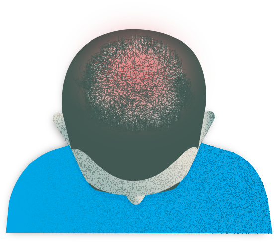
Telogen effluvium
What is telogen effluvium?
Telogen effluvium is a type of non-scarring alopecia that occurs when the hair follicle prematurely exits the growth (anagen) phase and enters the resting (telogen) phase, resulting in increased hair shedding.
It is one of the most common reasons for consultation, particularly in women, and can occur at any stage of life, as numerous triggers can disrupt the normal balance of the hair cycle.
Signs and symptoms of telogen effluvium
Diffuse, sudden and noticeable hair shedding is seen, typically 2-4 months after a triggering factor, resulting in a perceived overall reduction in hair volume without leading to completely bald areas.
Causes of telogen effluvium
Common triggers include infections, fever, surgery, periods of intense physical or emotional stress, hormonal changes—such as those occurring postpartum—restrictive diets, rapid weight loss, and nutritional deficiencies, particularly iron and vitamin D deficiency.
Diagnosis of telogen effluvium
Telogen effluvium is diagnosed through clinical examination by a specialist, based on observation of the scalp. Trichoscopy is used to confirm the diagnosis. This test allows assessment of variability in hair shaft thickness, an increased number of miniaturised follicles, reduced hair density and the presence of follicular units containing fewer hairs.
Treatment of telogen effluvium
Telogen effluvium is reversible and so has a good prognosis. Treatment focuses on identifying and addressing the underlying cause, as well as patient education, correction of associated deficiencies, and, in some cases, the use of medications to accelerate recovery and improve the regrowth phase.
Chemotherapy-induced anagen effluvium
What is anagen effluvium?
Anagen effluvium is a type of non-scarring alopecia and a common consequence of many oncological treatments. It occurs because hair follicle cells in the growth (anagen) phase have a high rate of division and are particularly sensitive to chemotherapeutic agents.
Signs and symptoms of anagen effluvium
It occurs as rapid, massive and generalised hair loss, which typically begins a few weeks after the onset of chemotherapy. Hair loss may be abrupt and affect other body hair, for example, the eyebrows and eyelashes, which can have a significant emotional impact on the patient.
Although its appearance can be alarming, in the vast majority of cases, the process is reversible, with progressive hair regrowth after completion of the treatment. Generally, this occurs within 3-6 months, although the rate and initial characteristics of regrowth may vary.
Causes of anagen effluvium
Key risk factors include the type of chemotherapy used, the cumulative dose and treatment cycle intensity. As a preventive measure, scalp cooling systems (cold caps) may reduce drug delivery to the follicle by decreasing local blood flow and, in some cases, lessen or delay hair loss. However, their effectiveness depends on the treatment regimen and they may not be suitable for all patients.
Treatment of anagen effluvium
Treatment focuses on emotional support, cosmetic measures and, in some cases, the use of medication to accelerate the regrowth phase.
Frontal fibrosing alopecia
What is frontal fibrosing alopecia?
Frontal fibrosing alopecia is a type of scarring alopecia characterised by a progressive, symmetrical and well-defined recession of the hairline along the forehead and temples. As it is a scarring alopecia, the lost hair does not regrow, making it crucial to seek medical advice at the first signs of eyebrow loss or frontal hairline recession, so that treatment can be started as early as possible.
It is currently one of the most common forms of scarring alopecia whose incidence has increased significantly in recent decades, particularly among postmenopausal women, although it may also affect premenopausal women and, more rarely, men.
Signs and symptoms of frontal fibrosing alopecia
The signs of frontal fibrosing alopecia include progressive, symmetrical recession of the frontal hairline. Partial or complete eyebrow loss is common and, in many cases, may be the first sign of the disease, even before it affects the scalp.
As the condition progresses, areas of alopecia may appear behind the ears or in other facial and body regions. Hair loss is often accompanied by symptoms such as itching (pruritus) or a sensation of burning or tightness.
Causes of frontal fibrosing alopecia
Although its exact cause is unknown, several hypotheses have been proposed without being conclusively confirmed. These include hormonal factors, immune system disturbances, the potential role of endocrine disruptors—whether through exposure to certain cosmetics or sunscreens—as well as environmental pollution.
Diagnosis of frontal fibrosing alopecia
Trichoscopy is essential for diagnosis, as it can identify possible signs of inflammation and follicular damage, including:
- Absence of follicular openings.
- Accumulation of keratin and inflammatory cells around the follicles.
- Vascular changes in the perifollicular area.
- Follicular inactivity.
Treatment of frontal fibrosing alopecia
Treatment aims to reduce inflammation and stabilise the disease through:
- Topical or injectable anti-inflammatory therapies to modulate the immune response.
- Topical treatments or medications (e.g., minoxidil) to improve hair thickness and density in unaffected areas.
- Topical and oral antiandrogens.
Other scarring alopecias
This group of alopecias includes disorders such as lichen planopilaris, folliculitis decalvans, cutaneous lupus erythematosus and central centrifugal cicatricial alopecia, which share a common outcome: irreversible destruction of the hair follicle.
Signs and symptoms of scarring alopecias
These alopecias occur as areas of hair loss accompanied by local inflammatory signs such as:
- Skin redness (erythema).
- Perifollicular scaling.
- Small pus-filled lesions (pustules).
- Pain or discomfort.
Although exact symptoms depend on the type of alopecia.
Over time, if inflammation persists, the skin in the affected area becomes smooth and thin, and the follicles disappear as scar tissue (fibrosis) forms. This is what prevents hair from regrowing.
Given their scarring nature, early detection is essential to prevent further permanent hair loss.
Causes of scarring alopecias
The causes are diverse and include immune system disorders, disturbances in the skin’s natural flora (microbiome), chronic infections, hormonal imbalances and genetic predisposition, although in many cases the exact cause remains unclear.
Diagnosis of scarring alopecias
Trichoscopy is a key tool for identifying characteristic findings such as:
- Absence of visible follicular openings.
- Accumulation of cells and keratin around the follicle (perifollicular hyperkeratosis).
- Skin debris and sebum obstructing the follicles (keratin plugs).
- Arborising blood vessels.
- Broken hairs or signs of inflammatory activity.
These findings help identify the specific subtype of alopecia and the degree of disease activity.
Treatment of scarring alopecias
As scarring alopecias, the lost hair does not regrow, making it crucial to seek medical advice at the first signs of eyebrow or hair loss, so treatment can be started as early as possible to prevent progression.
PortalClinic home
Joint project with
BBVA Foundation website.This link opens in a new tab.
Sorry this content wasn't helpful to you. Send us your comment and we will take it into account to continue improving.
Thanks for your help!
An error has occurred and we were unable to send your opinion, please try again later.
Substantiated information by:
Published: 13 May 2026
Updated: 13 May 2026
The donations that can be done through this webpage are exclusively for the benefit of Hospital Clínic of Barcelona through Fundació Clínic per a la Recerca Biomèdica and not for BBVA Foundation, entity that collaborates with the project of PortalClínic.
Subscribe
Receive the latest updates related to this content.
Thank you for subscribing!
If this is the first time you subscribe you will receive a confirmation email, check your inbox
An error occurred and we were unable to send your data, please try again later.